
Respiratory System
Enroll to start learning
You’ve not yet enrolled in this course. Please enroll for free to listen to audio lessons, classroom podcasts and take practice test.
Interactive Audio Lesson
Listen to a student-teacher conversation explaining the topic in a relatable way.
Anatomy of Airways and Alveoli
🔒 Unlock Audio Lesson
Sign up and enroll to listen to this audio lesson

Today, we will explore the respiratory system's anatomy. Can anyone tell me what the primary structures involved in respiration are?

The trachea and lungs?
Exactly! The trachea branches into bronchi and leads to the alveoli. Why do we need alveoli specifically?

To exchange gases?
That's right! The alveolar-capillary membrane is crucial for rapid gas diffusion. Remember, it's just 0.5 micrometers thick. Can someone summarize the difference between the upper and lower respiratory tracts?

The upper tract includes structures like nasal passages and the larynx, while the lower tract includes the trachea and alveoli.
Great summary! Let’s move on to the ventilation mechanisms.
Mechanics of Ventilation
🔒 Unlock Audio Lesson
Sign up and enroll to listen to this audio lesson

Can anyone describe what happens during inspiration?

The diaphragm contracts and moves downwards, increasing the thoracic volume.
Exactly! This leads to lower pressure in the thoracic cavity, allowing air to flow in. What about expiration?
It's mainly passive unless we forcefully exhale using internal intercostal muscles.
Correct! Remember, tidal volume is typically around 500 mL. Let's talk about lung volumes: can anyone name another lung capacity?
Inspiratory reserve volume?
Yes, that's about 3,000 mL! Understanding these volumes helps us assess lung function effectively.
Gas Exchange and Transport
🔒 Unlock Audio Lesson
Sign up and enroll to listen to this audio lesson
Let’s discuss gas exchange. How is oxygen transported in the blood?
Most of it is bound to hemoglobin, right?
Yes, about 98%! What's the significance of the oxyhemoglobin dissociation curve?
It shows how oxygen delivery changes with factors like pH and temperature.
Exactly! And how do we transport carbon dioxide?
It’s carried mostly as bicarbonate?
That's correct! Knowing these transport mechanisms is crucial for understanding respiratory physiology. Let’s summarize.
Respiratory Adaptations to Training
🔒 Unlock Audio Lesson
Sign up and enroll to listen to this audio lesson
What happens to our respiratory system with consistent training?
We have an increase in alveolar-capillary surface area!
That's right! What other adaptations might we expect?
Improved efficiency and a lower breathing frequency during exercise?
Exactly! These adaptations enhance performance. Understanding these can help us recognize the benefits of structured training.
Introduction & Overview
Read summaries of the section's main ideas at different levels of detail.
Quick Overview
Standard
This section details the structure of the respiratory system, including airways and alveoli, the mechanics of ventilation, gas exchange processes, and adaptations to training. Understanding these concepts is essential for optimizing physical performance and health.
Detailed
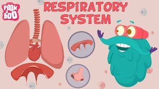
Respiratory System
The respiratory system plays a vital role in facilitating gas exchange between the body and the environment. It consists of the upper respiratory tract, including nasal passages, pharynx, and larynx, as well as the lower tract, which encompasses the trachea, bronchi, bronchioles, and alveoli. The thin alveolar-capillary membrane ensures efficient diffusion of gases.
Mechanics of ventilation involve two primary processes: inspiration and expiration. During inspiration, the diaphragm contracts and expands the thoracic cavity, resulting in decreased pressure and airflow into the lungs. Conversely, expiration is predominantly passive, involving relaxation of the diaphragm, although it may be aided by internal intercostal muscles for forced exhalation.
Lung volumes and capacities, such as tidal volume and vital capacity, are essential for assessing respiratory function. Gas exchange is facilitated by hemoglobin transport of oxygen and carbon dioxide, with parameters affected by the body's pH and temperature. Finally, the section discusses respiratory adaptations to training, including increased alveolar-capillary surface area and improved ventilatory efficiency, crucial for athletic performance.
Youtube Videos

Audio Book
Dive deep into the subject with an immersive audiobook experience.
Anatomy of Airways and Alveoli
Chapter 1 of 4
🔒 Unlock Audio Chapter
Sign up and enroll to access the full audio experience
Chapter Content
● Upper tract: nasal passages warm and filter; pharynx, larynx (voice production).
● Lower tract: trachea → bronchi → bronchioles → alveolar ducts → alveolar sacs.
● Alveolar-capillary membrane: thin barrier (~0.5 μm) for rapid gas diffusion.
Detailed Explanation
This chunk covers the anatomy of the respiratory system, specifically the structure of the airways and the alveoli. The upper tract consists of the nasal passages that have the dual role of warming and filtering the air we breathe, as well as the pharynx and larynx, which are involved in voice production. The lower tract is a pathway for air that includes the trachea, bronchi, and smaller branches called bronchioles, which lead to the alveolar ducts and finally to the alveolar sacs. The alveoli are tiny air sacs where gas exchange occurs and are surrounded by thin membranes (the alveolar-capillary membrane) that allow oxygen and carbon dioxide to diffuse quickly between the air and the bloodstream.
Examples & Analogies
Imagine blowing up a balloon. The trachea is like the neck of the balloon where air goes in, the bronchi are the main body that holds the air, and the alveoli are tiny balloons inside that help with the exchange of gases. Just like how air can easily fill a balloon, the thin alveolar membranes allow for easy transfer of oxygen and carbon dioxide.
Mechanics of Ventilation
Chapter 2 of 4
🔒 Unlock Audio Chapter
Sign up and enroll to access the full audio experience
Chapter Content
● Inspiration: diaphragm contracts (moves downward), external intercostals lift ribs; intrathoracic volume ↑, pressure ↓ → air inflow.
● Expiration: largely passive; diaphragm relaxes, internal intercostals for forced exhalation.
Lung volumes and capacities:
● Tidal volume (TV): ~500 mL
● Inspiratory reserve volume (IRV): ~3,000 mL
● Expiratory reserve volume (ERV): ~1,100 mL
● Residual volume (RV): ~1,200 mL
● Vital capacity (VC) = TV + IRV + ERV (~4,600 mL)
Detailed Explanation
This chunk explains how ventilation works – the process of inhaling and exhaling air. During inspiration, the diaphragm, which is a dome-shaped muscle at the base of the thoracic cavity, contracts and moves downward. This increase in volume in the chest cavity lowers the pressure, allowing air to flow into the lungs. During expiration, the diaphragm relaxes, and air is pushed out mostly passively. Some muscle action, via the internal intercostals, may be used if a forceful exhalation is needed. Key lung volumes include tidal volume (normal amount of air we breathe, around 500 mL), inspiratory reserve volume (extra air we can inhale, about 3,000 mL), expiratory reserve volume (extra air we can exhale, approximately 1,100 mL), and residual volume (air left in the lungs post-exhalation, around 1,200 mL). The total capability of the lungs to hold air is known as vital capacity.
Examples & Analogies
Think of your lungs like a balloon – when you take a deep breath in, the diaphragm pulls down like a balloon being filled with air, causing it to expand. When you let air out, it’s like when you let go of the balloon opening; most of the air comes out without much effort, but you can push even more out if you squeeze the balloon tightly.
Gas Exchange and Transport
Chapter 3 of 4
🔒 Unlock Audio Chapter
Sign up and enroll to access the full audio experience
Chapter Content
● Oxygen transport:
○ 98% bound to haemoglobin; 2% dissolved in plasma.
○ Oxyhaemoglobin dissociation curve: shifts with pH, temperature (Bohr effect).
● Carbon dioxide transport:
○ 70% as bicarbonate (HCO₃⁻) via carbonic anhydrase.
○ 23% bound to haemoglobin (carbaminohaemoglobin).
○ 7% dissolved.
Detailed Explanation
This section discusses how gases are transported in the body. Oxygen is primarily carried in the blood, with 98% bound to a protein called hemoglobin, while a small amount (2%) remains dissolved in plasma. The ability of hemoglobin to release oxygen to tissues depends on various factors, such as pH and temperature, which is explained by the oxyhemoglobin dissociation curve. In contrast, carbon dioxide transport is mainly done through bicarbonate ions (70%), while some attaches to hemoglobin (23%) and a smaller amount is dissolved in the blood (7%). This process is essential for maintaining the body's acid-base balance and for efficient respiration.
Examples & Analogies
Think of hemoglobin as a shuttle bus, picking up oxygen from the lungs and dropping it off at different stops (tissues) in the body. While most of the oxygen is carried as passengers in the bus, a few might just be standing near the bus (dissolved in plasma). For carbon dioxide, you can imagine it being collected from the body’s stops and taken back to the lungs to be released, similarly to bags of groceries you take home from the store.
Respiratory Adaptations to Training
Chapter 4 of 4
🔒 Unlock Audio Chapter
Sign up and enroll to access the full audio experience
Chapter Content
● Increased alveolar-capillary surface area.
● Enhanced pulmonary diffusion capacity.
● Improved ventilatory efficiency: lower breathing frequency at submaximal workloads.
Detailed Explanation
This segment highlights how the respiratory system adapts with regular training. With consistent exercise, the surface area available for gas exchange in the alveoli increases, which enhances the capacity for oxygen uptake and carbon dioxide release. Additionally, the efficiency of these processes improves so that during less intense workouts, people may find they don't need to breathe as rapidly or deeply to maintain adequate gas exchange. This leads to better overall respiratory fitness and endurance.
Examples & Analogies
Consider training your lungs like you would train your muscles. Just as lifting weights builds muscle bulk and strength, regular aerobic exercise helps expand the 'working area' of your lungs, making them more efficient. It's like upgrading from a small delivery truck to a big one that can carry more goods at once!
Key Concepts
-
Alveoli: Small air sacs essential for gas exchange.
-
Ventilation: The mechanics of breathing, including inspiration and expiration.
-
Gas Exchange: The process of oxygen and carbon dioxide transfer.
-
Training Adaptations: Changes in the respiratory system due to regular exercise.
Examples & Applications
Example 1: During physical exercise, the respiratory rate and tidal volume increase to meet higher oxygen demands.
Example 2: Athletes often exhibit improved pulmonary function due to enhanced alveolar-capillary surface area and ventilatory efficiency.
Memory Aids
Interactive tools to help you remember key concepts
Rhymes
When you take a breath in deep, your diaphragm moves and doesn’t sleep.
Memory Tools
For gas transport, remember: 'A B C' - Alveoli for binding, Blood for carrying, Cells for using.
Stories
Imagine your lungs as balloons. When you take a deep breath, they fill with air and expand, maximizing gas exchange at the alveoli. Expelling air is like letting air out of the balloon. Your diaphragm is the pump that makes it all possible.
Acronyms
V.A.C. = Ventilation, Alveoli, Capacity to remember parts of respiration.
Flash Cards
Glossary
- Alveoli
Small air sacs in the lungs where gas exchange occurs.
- Tidal Volume
The amount of air inhaled or exhaled during normal breathing, approximately 500 mL.
- Inspiratory Reserve Volume (IRV)
The maximum amount of additional air that can be inhaled after a normal breath, around 3,000 mL.
- Residual Volume (RV)
The volume of air remaining in the lungs after maximum exhalation, about 1,200 mL.
- Gas Exchange
The process of exchanging oxygen and carbon dioxide between the alveoli and bloodstream.
- Bohr Effect
The physiological phenomenon where hemoglobin's oxygen binding affinity decreases as pH drops.
Reference links
Supplementary resources to enhance your learning experience.