
Human Respiratory System
Enroll to start learning
You’ve not yet enrolled in this course. Please enroll for free to listen to audio lessons, classroom podcasts and take practice test.
Interactive Audio Lesson
Listen to a student-teacher conversation explaining the topic in a relatable way.
Overview of the Human Respiratory System
🔒 Unlock Audio Lesson
Sign up and enroll to listen to this audio lesson

Today, we're going to learn about the human respiratory system. Can anyone tell me the main functions of this system?

It helps us breathe in oxygen and release carbon dioxide.
Exactly! The respiratory system not only supplies oxygen but is also critical in expelling carbon dioxide from our bodies. Let’s start with its structure. What do you think the respiratory system includes?

The lungs, I think?
Yes! The lungs are the main organs, but they work with several other structures like the nostrils, nasal cavity, pharynx, and trachea. Remember, we can use the acronym 'NLPTAB' — Nostrils, Larynx, Pharynx, Trachea, Alveoli, Bronchi — to help us remember these key components.

How do these parts actually work together?
Great question! Each part plays a role in filtering, humidifying, and directing the air. The trachea, for instance, divides into bronchi, leading into bronchioles, all eventually connecting to the alveoli where gas exchange happens.

So, it’s like a highway leading to a delivery area?
Nicely put! The alveoli are indeed the delivery area for oxygen. Let's summarize: the respiratory system consists of several organs that work together to facilitate breathing and gas exchange.
Mechanism of Breathing
🔒 Unlock Audio Lesson
Sign up and enroll to listen to this audio lesson

Now, let’s discuss how we breathe. What happens during inspiration?
The diaphragm contracts and pulls down, which makes the chest cavity larger.
And this creates lower pressure inside the lungs compared to the outside air, so air flows in!
Exactly! During expiration, what changes occur?
The diaphragm relaxes, which pushes the air out because the pressure inside increases?
Spot on! The diaphragm moves back up, and the intercostal muscles also help change the volume of the thorax, prompting air to move out. Remember the mnemonic 'CRYPT' – Contract Relax, Yield Pressure for the breathing mechanism!
How do we measure how much air we breathe?
That's where spirometry comes in! It measures different volumes of air, like tidal volume and expiratory reserve volume, helping assess lung function.
It sounds vital for diagnosing respiratory conditions!
Absolutely! Understanding the mechanism of breathing is essential for everyone who studies biology, especially human health. Let's summarize: breathing involves creating pressure changes through diaphragm and rib cage movement.
Gas Exchange and Transport in the Body
🔒 Unlock Audio Lesson
Sign up and enroll to listen to this audio lesson
Next, we focus on the exchange of gases. Can anyone explain where gas exchange occurs?
In the alveoli, right?
Yes! The alveoli are the sites for the diffusion of oxygen and carbon dioxide. Why do you think this process happens in the alveoli?
Because they are thin and have a large surface area for better diffusion?
Exactly! The structure of the alveoli maximizes efficiency. Now, how does oxygen move from the alveoli to our blood?
It moves from a higher concentration in the alveoli to a lower concentration in the blood.
Right! This process is driven by concentration gradients, and the same goes for carbon dioxide moving in the opposite direction. Can you remember the terms 'partial pressure' used for these gases?
Yes! It's important for understanding how gases behave.
Exactly! And once in the blood, oxygen is primarily transported by hemoglobin. Let's wrap up: gas exchange and transport are essential functions of the respiratory system, relying on diffusion and pressure gradients.
Introduction & Overview
Read summaries of the section's main ideas at different levels of detail.
Quick Overview
Standard
The human respiratory system is essential for the intake of oxygen and the expulsion of carbon dioxide through a series of interconnected organs, including the nostrils, nasal chamber, pharynx, larynx, trachea, bronchi, and lungs. It operates in two main phases: inspiration and expiration, supported by muscle movements to create pressure gradients necessary for breathing.
Detailed
Human Respiratory System
The human respiratory system is composed of a set of organs responsible for breathing and gas exchange. It begins with external nostrils located above the upper lip and leads into the nasal passages, where air is filtered and humidified. The air then moves into the pharynx, a shared passageway for food and air, and progresses to the larynx, known as the sound box due to its role in sound production. The trachea extends downwards and divides into the right and left primary bronchi, which become progressively smaller and branch out into bronchioles, eventually leading to alveoli—tiny air sacs where gas exchange occurs.
Breathing, or pulmonary ventilation, consists of two phases: inspiration (air intake) and expiration (air expulsion). The diaphragm and intercostal muscles work together to create pressure changes within the thoracic cavity, allowing air to flow into the lungs during inspiration and forcing it out during expiration.
Respiration also involves crucial processes like gas exchange, where oxygen diffuses from the alveoli into the blood, and carbon dioxide diffuses in the opposite direction. Additionally, gases are transported through the bloodstream—most oxygen binds with hemoglobin in red blood cells, while carbon dioxide is transported in various forms, including dissolved in plasma.
The anatomical setup within the thoracic cavity and the function of respiratory organs are vital for maintaining effective breathing and ensuring that oxygen reaches tissues while removing carbon dioxide effectively.
Youtube Videos


Audio Book
Dive deep into the subject with an immersive audiobook experience.
Structure of the Human Respiratory System
Chapter 1 of 6
🔒 Unlock Audio Chapter
Sign up and enroll to access the full audio experience
Chapter Content
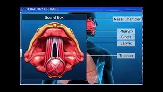
We have a pair of external nostrils opening out above the upper lips. It leads to a nasal chamber through the nasal passage. The nasal chamber opens into the pharynx, a portion of which is the common passage for food and air. The pharynx opens through the larynx region into the trachea.
Detailed Explanation
The structure of the human respiratory system starts with the external nostrils located above the upper lip. These nostrils lead into the nasal chamber via the nasal passage. The nasal chamber is important for filtering and warming the air we breathe. It opens into the pharynx, which serves as a common pathway for both food entering the esophagus and air heading to the lungs. The pharynx is connected to the larynx, a pivotal organ for sound production, which leads into the trachea, the main airway to the lungs.
Examples & Analogies
Think of your respiratory system like a pathway in a park. The nostrils are like the park gates where people enter. Once they pass through the gates (nostrils), they walk through a nice, sheltered path (nasal chamber) that leads to various areas in the park (pharynx), eventually arriving at different sections of the park dedicated for activities, just like air is directed to the lungs.
Larynx and Trachea Functions
Chapter 2 of 6
🔒 Unlock Audio Chapter
Sign up and enroll to access the full audio experience
Chapter Content
Larynx is a cartilaginous box which helps in sound production and hence called the sound box. During swallowing glottis can be covered by a thin elastic cartilaginous flap called epiglottis to prevent the entry of food into the larynx. Trachea is a straight tube extending up to the mid-thoracic cavity, which divides at the level of 5th thoracic vertebra into a right and left primary bronchi.
Detailed Explanation
The larynx, often referred to as the 'voice box', plays a crucial role in sound production; it houses the vocal cords. To prevent food from entering the trachea when swallowing, a flap called the epiglottis covers the opening of the larynx. The trachea is a straight tube that serves as the main airway, extending down to the thoracic cavity and branching into two primary bronchi that lead into each lung.
Examples & Analogies
Imagine the larynx as the speaker in a concert, creating sound that can be heard by everyone. The epiglottis acts like a lid on a container: when you're about to take a drink (swallow), it closes over the opening to make sure nothing but the liquid goes in, not solid food.
Bronchi and Alveoli Structure
Chapter 3 of 6
🔒 Unlock Audio Chapter
Sign up and enroll to access the full audio experience
Chapter Content
Each bronchi undergoes repeated divisions to form the secondary and tertiary bronchi and bronchioles ending up in very thin terminal bronchioles. Each terminal bronchiole gives rise to a number of very thin, irregular-walled and vascularised bag-like structures called alveoli.
Detailed Explanation
After the trachea divides into the primary bronchi, these bronchi continue to branch into smaller bronchi and ultimately into tiny terminal bronchioles, which are the last segments of the bronchi. From each terminal bronchiole, a network of tiny structures called alveoli forms. Alveoli are crucial for gas exchange; they are thin-walled and surrounded by blood vessels, allowing oxygen and carbon dioxide to be exchanged efficiently.
Examples & Analogies
Picture the branching roads of a city that lead to smaller streets; the bronchi serve as the main roads, while bronchioles and alveoli are like side streets where the smaller interactions take place — in this case, the vital exchange of gases that happens at the alveoli.
Lung Structure and Pleura
Chapter 4 of 6
🔒 Unlock Audio Chapter
Sign up and enroll to access the full audio experience
Chapter Content
We have two lungs which are covered by a double layered pleura, with pleural fluid between them. It reduces friction on the lung-surface. The outer pleural membrane is in close contact with the thoracic lining whereas the inner pleural membrane is in contact with the lung surface.
Detailed Explanation
The human body contains two lungs, and each lung is enveloped in a double-layered membrane known as the pleura. The space between these two layers contains pleural fluid, which allows the lungs to move smoothly against the thoracic cavity wall during breathing, minimizing friction. The outer layer adheres to the chest wall while the inner layer closely wraps around the lung itself.
Examples & Analogies
Think of the pleura as a protective glove for each lung. Just like a glove helps your hand move smoothly and freely, the pleura allows the lungs to expand and contract effortlessly without getting damaged from friction.
Function of Respiratory System Parts
Chapter 5 of 6
🔒 Unlock Audio Chapter
Sign up and enroll to access the full audio experience
Chapter Content
The part starting with the external nostrils up to the terminal bronchioles constitute the conducting part whereas the alveoli and their ducts form the respiratory or exchange part of the respiratory system.
Detailed Explanation
The entire structure of the human respiratory system can be categorized into two parts: the conducting part and the respiratory part. The conducting part encompasses the external nostrils to the terminal bronchioles; its main function is to transport air while also filtering, warming, and humidifying it. The respiratory part consists of the alveoli and their ducts, where the actual gas exchange occurs between air in the alveoli and the blood in capillaries.
Examples & Analogies
Consider the conducting part as the delivery route to a restaurant where food is prepared (alveoli). The roads serve to transport customers comfortably and safely to their dining experience, just as air is prepared for the lungs' gas exchange.
Breathing Process
Chapter 6 of 6
🔒 Unlock Audio Chapter
Sign up and enroll to access the full audio experience
Chapter Content
The lungs are situated in the thoracic chamber which is anatomically an air-tight chamber. The thoracic chamber is formed dorsally by the vertebral column, ventrally by the sternum, laterally by the ribs and on the lower side by the dome-shaped diaphragm.
Detailed Explanation
The lungs reside within the thoracic cavity, a sealed space that protects the lungs and facilitates their function. The thoracic cavity is bounded by the vertebral column at the back, the sternum (breastbone) at the front, ribs forming the sides, and the diaphragm, which is a dome-shaped muscle located beneath the lungs. This setup is crucial for effective breathing because as the volume of this cavity changes, it influences the lungs' volume and the air pressure within them.
Examples & Analogies
Imagine the thoracic cavity as a balloon. When you squeeze the sides of the balloon (similar to contracting the diaphragm), it inflates and expands (the lungs fill with air) while releasing air when you let go. This function enables us to breathe in and out effectively.
Key Concepts
-
Breathing: The process by which air is inhaled (inspiration) and exhaled (expiration).
-
Gas Exchange: The diffusion of oxygen into the blood and carbon dioxide out of the blood at the alveoli.
-
Transport of Gases: How oxygen is carried in the blood primarily through hemoglobin, while carbon dioxide is transported in different forms including bicarbonate.
Examples & Applications
When a person inhales, atmospheric pressure is greater than the pressure in the lungs, resulting in air rushes into the lungs.
During exercise, the respiratory rate and depth of breathing increase to meet the higher oxygen demands of the body.
Memory Aids
Interactive tools to help you remember key concepts
Rhymes
Breathe in with ease, the air we seize, through nostrils it flows, to lungs it goes.
Stories
Imagine your lungs as balloons that fill with air when you inhale through a straw. When you breathe out, they deflate, pushing the air back through the straw.
Memory Tools
Remember the diaphragm’s role: It 'drops' for a deep breath and 'relaxes' to let out air.
Acronyms
Use 'LPNTR' for lungs, pharynx, nasal cavity, trachea, and respiratory pathways.
Flash Cards
Glossary
- Respiratory System
The organ system responsible for breathing, gas exchange, and oxygen transport.
- Alveoli
Tiny air sacs in the lungs where gas exchange occurs.
- Hemoglobin
An iron-containing protein in red blood cells that binds to oxygen for transport.
- Inspiration
The process of drawing air into the lungs.
- Expiration
The process of expelling air from the lungs.
- Diffusion
The movement of gases from areas of high concentration to areas of low concentration.
- Spirometry
A technique used to measure lung volumes and capacities.
Reference links
Supplementary resources to enhance your learning experience.