
Mechanism of Muscle Contraction
Enroll to start learning
You’ve not yet enrolled in this course. Please enroll for free to listen to audio lessons, classroom podcasts and take practice test.
Interactive Audio Lesson
Listen to a student-teacher conversation explaining the topic in a relatable way.
Introduction to Muscle Contraction
🔒 Unlock Audio Lesson
Sign up and enroll to listen to this audio lesson

Today, we're discussing the mechanism of muscle contraction, specifically the sliding filament theory. Who can tell me what initiates this process?

Is it a signal from the nervous system?
Exactly! The contraction starts with a signal from the central nervous system via a motor neuron. This signal reaches the neuromuscular junction. Can anyone explain what happens at this junction?

That's where Acetylcholine is released, right?
Correct! The release of Acetylcholine generates an action potential in the muscle fiber, leading to contraction. Remember the acronym 'ANAP' for Action potential, Nuromuscular junction, Acetylcholine, and Potential – it highlights the initiation steps! Let's move to what happens next.
Calcium's Role in Contraction
🔒 Unlock Audio Lesson
Sign up and enroll to listen to this audio lesson

Once the action potential spreads, what occurs next in the muscle fiber?

The sarcoplasmic reticulum releases calcium ions!
Right! The rise of calcium levels triggers binding to troponin, allowing myosin heads to attach to actin. This change is key for the sliding mechanism. How does this binding occur?

The active sites on actin are exposed, allowing myosin to bind.
Perfect! So remember: calcium is like a key that unlocks the actin for myosin interaction. We can say 'Ca²⁺ unlocks action' to remember calcium's role!
Cross-Bridge Cycle
🔒 Unlock Audio Lesson
Sign up and enroll to listen to this audio lesson
Now, let's discuss the interaction between myosin and actin in detail. What happens when myosin heads bind to actin?
The myosin pulls the actin filaments toward the center, causing contraction.
Exactly! This is known as the cross-bridge cycle. Who can outline the steps involved in this cycle causing contraction?
First, the myosin head binds to actin, then it pulls it inward, and then it releases and resets with ATP.
Great overview! Let's create the mnemonic 'BPR - Bind, Pull, Reset' to summarize these actions. As you can see, muscle contraction is all about the rhythmic binding and releasing of these filaments!
Muscle Relaxation Process
🔒 Unlock Audio Lesson
Sign up and enroll to listen to this audio lesson
After muscle contraction, we need to understand how relaxation occurs. What role does calcium play here?
Calcium ions are pumped back into the sarcoplasmic reticulum.
Correct. This allows the active sites on actin to be re-masked by tropomyosin. Why is this significant?
It leads the muscle to return to its original length—muscle relaxation!
Excellent understanding! To remember, think: 'Calcium out, muscle relaxes.' This process is just as important as contraction!
Fatigue in Muscle Contraction
🔒 Unlock Audio Lesson
Sign up and enroll to listen to this audio lesson
What happens to muscles when they contract repeatedly for a long time?
They get fatigued?
Correct! Fatigue can occur due to lactic acid accumulation from anaerobic respiration. Can anyone state how we can tell apart aerobic muscles from anaerobic ones?
Aerobic muscles have more myoglobin and rely on oxygen, while anaerobic muscles are paler and have very little myoglobin.
Exactly! Remember: 'Red is for aerobic' for muscles rich in myoglobin, and they’re better for endurance than the pale anaerobic muscles. This knowledge is crucial for understanding muscle training.
Introduction & Overview
Read summaries of the section's main ideas at different levels of detail.
Quick Overview
Standard
Muscle contraction occurs through the sliding filament theory, initiated by signals from the central nervous system that activate motor neurons. The binding of calcium ions to muscle proteins facilitates the interaction between actin and myosin, leading to contraction.
Detailed
Mechanism of Muscle Contraction
Muscle contraction is crucial for movement, and it is explained by the sliding filament theory. This theory proposes that muscle fibers contract through the sliding motion of thin (actin) filaments over thick (myosin) filaments.
Process of Contraction:
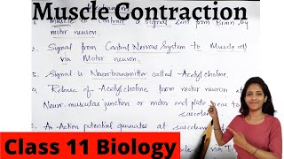
- Initiation: The process is initiated by a neural signal from the central nervous system (CNS) traveling through a motor neuron to the junction known as the neuromuscular junction or motor-end plate.
- When the signal reaches this junction, it stimulates the release of a neurotransmitter called Acetylcholine.
- Action Potential: The release of Acetylcholine generates an action potential in the sarcolemma (muscle cell membrane) which quickly spreads throughout the muscle fiber.
- Calcium Release: The action potential causes the sarcoplasmic reticulum (specialized endoplasmic reticulum in muscle cells) to release calcium ions (Ca²⁺) into the sarcoplasm (muscle cell cytoplasm).
- Tropomyosin and Troponin Interaction: The increase in calcium ions interacts with troponin, a protein associated with the actin filaments, leading to the exposure of active sites on actin that allow myosin to bind.
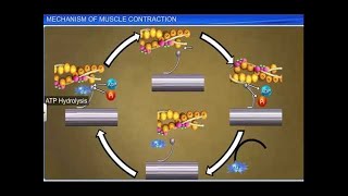
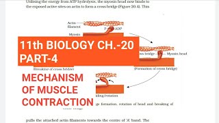
- Cross-Bridge Formation: The myosin heads, energized by ATP hydrolysis, bind to the exposed active sites on actin, forming a cross-bridge.
- Sliding Mechanism: The myosin head pulls the actin filaments toward the center of the sarcomere (the functional unit of muscle), resulting in muscle contraction. The myosin head returns to a relaxed state after releasing ADP and inorganic phosphate (Pi), and a new ATP molecule binds to the myosin head, breaking the cross-bridge.
- Relaxation: As Ca²⁺ ions are pumped back into the sarcoplasmic reticulum, the actin sites are re-masked by tropomyosin, leading the muscle to relax back to its original length.
This cycle continues as long as calcium ions remain high in concentration, allowing for muscle contractions until fatigue occurs, often characterized by the accumulation of lactic acid.
Youtube Videos


Audio Book
Dive deep into the subject with an immersive audiobook experience.
Sliding Filament Theory
Chapter 1 of 8
🔒 Unlock Audio Chapter
Sign up and enroll to access the full audio experience
Chapter Content
Mechanism of muscle contraction is best explained by the sliding filament theory which states that contraction of a muscle fibre takes place by the sliding of the thin filaments over the thick filaments.
Detailed Explanation
The sliding filament theory explains how muscles contract. According to this theory, muscle contraction occurs when the thin actin filaments slide over the thick myosin filaments within muscle fibers. This sliding is what shortens the muscle and produces movement.
Examples & Analogies
Imagine a pair of scissors. When you close them, the blades slide past one another to cut. Similarly, when muscle fibers contract, the actin filaments slide over the myosin filaments, bringing about movement.
Initiation of Muscle Contraction
Chapter 2 of 8
🔒 Unlock Audio Chapter
Sign up and enroll to access the full audio experience
Chapter Content
Muscle contraction is initiated by a signal sent by the central nervous system (CNS) via a motor neuron. A motor neuron alongwith the muscle fibres connected to it constitute a motor unit. The junction between a motor neuron and the sarcolemma of the muscle fibre is called the neuromuscular junction or motor-end plate.
Detailed Explanation
Muscle contraction begins when the brain sends a signal through motor neurons. These neurons connect to muscle fibers at a junction called the neuromuscular junction. This connection is crucial because it is where the electrical signal from the neuron triggers the muscle fiber to begin contracting.
Examples & Analogies
Think of a remote control car. When you press a button on the remote, a signal is sent to the car to move. Similarly, when the CNS sends a signal to the motor neuron, it tells the muscle to contract.
Role of Neurotransmitters
Chapter 3 of 8
🔒 Unlock Audio Chapter
Sign up and enroll to access the full audio experience
Chapter Content
A neural signal reaching this junction releases a neurotransmitter (Acetyl choline) which generates an action potential in the sarcolemma. This spreads through the muscle fibre and causes the release of calcium ions into the sarcoplasm.
Detailed Explanation
When the nerve impulse reaches the neuromuscular junction, it releases a neurotransmitter called acetylcholine. This chemical messengers generate an action potential or electrical change in the muscle fiber, which causes calcium ions to be released from storage within the muscle cell. Calcium is essential for muscle contraction as it activates the proteins that enable the muscles to slide and contract.
Examples & Analogies
Consider a water balloon. When you squeeze a balloon, it causes the water inside to move. Similarly, the release of acetylcholine at the neuromuscular junction causes calcium ions to flood the sarcoplasm, initiating the contraction process.
Cross Bridge Formation
Chapter 4 of 8
🔒 Unlock Audio Chapter
Sign up and enroll to access the full audio experience
Chapter Content
Increase in Ca++ level leads to the binding of calcium with a subunit of troponin on actin filaments and thereby remove the masking of active sites for myosin. Utilising the energy from ATP hydrolysis, the myosin head now binds to the exposed active sites on actin to form a cross bridge.
Detailed Explanation
When calcium ions increase, they bind to troponin, which changes shape and exposes binding sites on actin filaments. The myosin heads, energized by ATP, attach to these sites to form cross bridges. This attachment is crucial for the sliding movement that results in contraction.
Examples & Analogies
Imagine a person using magnets to pick up metal objects. The magnets can only grab onto the metal when they are close enough. Similarly, myosin heads can only bind to actin when the actin's binding sites are uncovered.
Muscle Shortening
Chapter 5 of 8
🔒 Unlock Audio Chapter
Sign up and enroll to access the full audio experience
Chapter Content
This pulls the attached actin filaments towards the centre of ‘A’ band. The ‘Z’ line attached to these actins are also pulled inwards thereby causing a shortening of the sarcomere, i.e., contraction.
Detailed Explanation
When the cross bridges form and rotate, they pull the actin filaments closer together. This action draws the Z lines closer, which shortens the sarcomere—the functional unit of muscle contraction. Ultimately, this shortening of the sarcomeres leads to the overall contraction of the muscle.
Examples & Analogies
Think of a rubber band. If you pull on both sides, the band stretches. But when you let go, it contracts back to its original size. In muscle fibers, contraction pulls the actin filaments inward.
Muscle Relaxation
Chapter 6 of 8
🔒 Unlock Audio Chapter
Sign up and enroll to access the full audio experience
Chapter Content
The myosin, releasing the ADP and P goes back to its relaxed state. A new ATP binds and the cross-bridge is broken. The ATP is again hydrolysed by the myosin head and the cycle of cross bridge formation and breakage is repeated causing further sliding.
Detailed Explanation
After the power stroke, the myosin head releases ADP and phosphate, reverting to its original position. A new ATP molecule then binds to the myosin, causing it to detach from actin, breaking the cross bridge. This cycle can repeat as long as ATP and calcium are present, allowing for continued muscle contraction.
Examples & Analogies
Like a continuous loop in a conveyor belt, as long as there is energy (ATP) available, the myosin heads can repeatedly pull actin filaments, facilitating muscle contraction.
Fatigue and Recovery
Chapter 7 of 8
🔒 Unlock Audio Chapter
Sign up and enroll to access the full audio experience
Chapter Content
Repeated activation of the muscles can lead to the accumulation of lactic acid due to anaerobic breakdown of glycogen in them, causing fatigue. Muscle contains a red coloured oxygen storing pigment called myoglobin.
Detailed Explanation
When muscles are used intensively over a period, they can become fatigued due to lactic acid buildup from anaerobic respiration. Myoglobin in muscle cells helps to store oxygen, which is crucial for aerobic respiration, helping muscles recover and maintain energy levels.
Examples & Analogies
Think of running a marathon. As you push your body, it starts getting tired due to lack of oxygen, similar to how muscles produce lactic acid when they run out of oxygen during intense activity.
Red vs. White Muscle Fibers
Chapter 8 of 8
🔒 Unlock Audio Chapter
Sign up and enroll to access the full audio experience
Chapter Content
Muscles with high myoglobin content are called Red fibres. These muscles also contain plenty of mitochondria which can utilise the large amount of oxygen stored in them for ATP production. These muscles, therefore, can also be called aerobic muscles. On the other hand, some muscles possess very less quantity of myoglobin and therefore, appear pale or whitish. These are the White fibres.
Detailed Explanation
Muscle fibers can be categorized into red fibers and white fibers based on their myoglobin content. Red fibers, which are rich in myoglobin and mitochondria, are designed for endurance and aerobic activities, while white fibers, with lower myoglobin, perform better in short, quick bursts of strength, relying more on anaerobic metabolism.
Examples & Analogies
Think of red fibers as marathon runners and white fibers as sprinters. The runners (red fibers) build endurance over long distances, while sprinters (white fibers) excel in short, high-intensity bursts.
Key Concepts
-
Sliding Filament Theory: Explains how muscle contraction occurs as thin filaments slide over thick filaments.
-
Neuromuscular Junction: The point where a motor neuron communicates with a muscle fiber to initiate contraction.
-
Calcium Ions Role: Calcium ions bind to troponin, exposing active sites on actin for myosin binding.
Examples & Applications
An example of sliding filament theory is seen when lifting weights; as the muscle contracts, actin and myosin filaments slide past each other.
The fatigue experienced after intense exercise is due to the buildup of lactic acid resulting from prolonged muscle contraction.
Memory Aids
Interactive tools to help you remember key concepts
Rhymes
To lift, to pull, to slide with grace, Actin and Myosin in their place.
Stories
Imagine a key (calcium) that unlocks a door (tropomyosin) to a room (active sites) where myosin is waiting to dance with actin, sliding them closer every beat!
Memory Tools
The acronym 'CAP' can help remember the process: Calcium release, Actin binding, Power stroke for contraction.
Acronyms
Use 'CALMS' for remembering muscle contraction steps
for Calcium
for Actin
for Link
for Move
for Slide.
Flash Cards
Glossary
- Sliding filament theory
A theory explaining muscle contraction through the sliding of actin and myosin filaments.
- Neuromuscular junction
The synapse between a motor neuron and a muscle fiber.
- Acetylcholine
A neurotransmitter released at the neuromuscular junction.
- Sarcoplasmic reticulum
The specialized endoplasmic reticulum in muscle fibers that stores calcium ions.
- Crossbridge
The connection formed between myosin and actin filaments during contraction.
Reference links
Supplementary resources to enhance your learning experience.