
Monitoring and Measurement of Microorganisms
Enroll to start learning
You’ve not yet enrolled in this course. Please enroll for free to listen to audio lessons, classroom podcasts and take practice test.
Interactive Audio Lesson
Listen to a student-teacher conversation explaining the topic in a relatable way.
Introduction to Microorganism Measurement Standards
🔒 Unlock Audio Lesson
Sign up and enroll to listen to this audio lesson

Today we're discussing the monitoring standards for microorganisms in water, specifically focusing on how we determine acceptable levels of pathogens. Can anyone tell me what the CPCB standard is for microorganisms in drinking water?

Is it five microorganisms per 100 ml, sir?
Exactly! This means that any water should ideally contain no more than five pathogens in a 100 ml sample. Why do you think it's important to monitor these levels?

To ensure that the water is safe for human consumption and to prevent diseases.
Right! Monitoring pathogens helps prevent infections. Now, let's understand how we actually count these microorganisms.
Counting Techniques: The Filtration Method
🔒 Unlock Audio Lesson
Sign up and enroll to listen to this audio lesson

One key method for counting microorganisms is filtration. What do you think filtering does?

It isolates the microorganisms so we can see them better, right?
Exactly! By filtering a 100 ml sample of water, we can catch the microbes on a filter paper. Then we can examine it under a microscope. What challenges do you think might arise with this method?

Finding the tiny colonies on the filter could be really tough!
Correct! Each colony is very small, which makes it challenging to distinguish them. This necessitates using other methods for better clarity.
Culturing Microorganisms
🔒 Unlock Audio Lesson
Sign up and enroll to listen to this audio lesson
Let's discuss the culturing method. How does this method help in counting microorganisms?
It allows them to grow into larger colonies that we can see and count!
Exactly! By placing a water sample on a nutrient medium and incubating it, we can stimulate the growth of microorganisms into colonies. What do we call these clusters of bacteria?
Colony-forming units or CFUs!
Well done! Remember, if the concentration is too high, we might need to dilute the sample first to get distinct CFUs.
Advanced Techniques: Flow Cytometry and Staining
🔒 Unlock Audio Lesson
Sign up and enroll to listen to this audio lesson
Moving beyond traditional methods, flow cytometry is a fascinating technique used now. Can anyone explain how it works?
It counts cells one at a time as they flow through a channel, right?
That's correct! However, it's not widely adopted yet due to issues like sample representativeness. Now, let's talk about staining. How does staining improve our ability to observe microorganisms?
It helps differentiate between types of bacteria, especially if we use fluorescent dyes.
Exactly! Staining enhances visibility under a microscope. Both flow cytometry and staining are valuable, but traditional culturing remains a cornerstone for microbial analysis.
Introduction & Overview
Read summaries of the section's main ideas at different levels of detail.
Quick Overview
Standard
Monitoring microorganisms, particularly pathogens in water, is crucial for maintaining environmental quality. The section discusses various analytical methods, including culturing, microscopy, and flow cytometry, while addressing challenges like accurate counting and the need for dilution in high concentrations.
Detailed
Monitoring and Measurement of Microorganisms
This section delves into the significance of monitoring microorganisms, especially pathogens, to ensure water quality. The Central Pollution Control Board (CPCB) sets standards for acceptable levels of microbes, typically five per 100 ml in drinking water. Given the microscopic size of bacteria (1-10 microns), direct counting in water samples is challenging. Therefore, a common method employs filtration to isolate microorganisms on a filter paper, which is then examined under a microscope.
A fundamental approach to assessing microbial populations is the culturing method. In this method, a water sample is introduced into a nutrient medium that fosters bacterial growth. After incubation for 24 hours at a controlled temperature, colonies form, which can be counted as colony-forming units (CFU). High concentrations of microorganisms lead to complications in differentiation; thus, dilution before culturing is often necessary to yield distinct colonies.
In addition to culturing, alternative techniques such as flow cytometry—which counts particles in liquid samples—are emerging but have yet to be standardized in routine microbial analysis. Other methodologies include staining to visualize bacteria under fluorescence microscopy, as well as advanced DNA analysis for accurate identification. The presence of turbidity often indicates high microbial concentration, prompting further examination for viability.
This section emphasizes that the ability to distinguish viable microorganisms is critical in assessing potential health risks, ultimately determining water safety.
Youtube Videos


Audio Book
Dive deep into the subject with an immersive audiobook experience.
Standards for Microorganism Analysis
Chapter 1 of 9
🔒 Unlock Audio Chapter
Sign up and enroll to access the full audio experience
Chapter Content
So, here if we look at the standards that people use for analysis of microorganisms, for example CPCB standards for microorganisms will be like say 5 per 100 ml or 5 microorganisms for 100 ml. So, the microorganisms we are talking about are predominantly pathogens. Many of the pathogens are bacteria, there are a few viruses and all that but mainly bacteria and for water quality, people count the number of bacteria in it.
Detailed Explanation
In this section, we learn about the standards set for counting microorganisms, particularly in water. The Central Pollution Control Board (CPCB) has established guidelines stating that for water quality analysis, there should be at most 5 pathogenic microorganisms per 100 milliliters of water. Pathogens are harmful microorganisms, primarily bacteria, which can cause diseases. Therefore, monitoring these levels is crucial for ensuring safe water quality.
Examples & Analogies
Imagine checking your drinking water for safety like you would check the amount of salt when cooking. Just as you wouldn't want too much salt in your food, you want to ensure that the water has a safe level of harmful microorganisms.
Counting Microorganisms
Chapter 2 of 9
🔒 Unlock Audio Chapter
Sign up and enroll to access the full audio experience
Chapter Content
So, how do you count say 5 per 100 ml? This bacteria size is around 1 to 10 microns. It means if I take 100 ml sample, I have to see it, it is difficult to count, so you need a microscope.
Detailed Explanation
Counting microorganisms is challenging because they are very small, typically between 1 to 10 microns in size. To count them, a 100 ml water sample is needed, but direct counting with the naked eye is impossible. Therefore, a microscope is necessary to observe and count these tiny organisms accurately.
Examples & Analogies
Think of trying to count grains of sand on a beach. It's nearly impossible to do it by eye because they are so tiny. Just as a magnifying glass can help you see the sand better, a microscope helps scientists see and count microorganisms.
Culturing Method for Microorganism Counting
Chapter 3 of 9
🔒 Unlock Audio Chapter
Sign up and enroll to access the full audio experience
Chapter Content
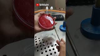
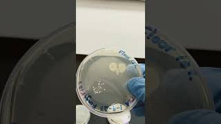
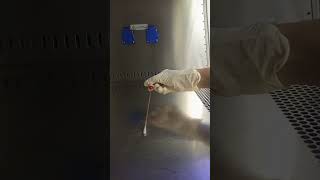
One of the old standard methods is that people use what is called a culturing method. ...you take a plate filled with some nutrients. ...you take say 1 ml of water sample and put it on a nutrient medium.
Detailed Explanation
An established method for counting microorganisms is the culturing method. In this process, a water sample (1 ml) is placed on a nutrient-rich medium in a petri dish. The nutrients allow bacteria to grow and multiply. After incubating for 24 hours at a specific temperature, bacterial colonies form, making it easier to count them.
Examples & Analogies
It's similar to baking bread. You mix ingredients to create the right environment for yeast to grow and rise. Just as the yeast multiplies in the dough, bacteria multiply in the nutrient medium, forming visible colonies.
Counting Colony Forming Units (CFUs)
Chapter 4 of 9
🔒 Unlock Audio Chapter
Sign up and enroll to access the full audio experience
Chapter Content
So, this is called as CFU or a colony forming unit. ...So therefore, you cannot get an immediate value of this thing...
Detailed Explanation
The results of culturing are expressed as Colony Forming Units (CFUs). This refers to the number of visible colonies that grow from one original bacterial cell. Since incubation takes time, it is impossible to get instant results. After waiting, scientists can count the colonies formed and relate this back to the volume of the original sample.
Examples & Analogies
Imagine planting seeds in a garden. You can't see how many plants will grow right away; you must wait for them to sprout and develop. In the same way, the bacteria must be given time to grow into colonies before counting them.
Dilution for High Concentration Samples
Chapter 5 of 9
🔒 Unlock Audio Chapter
Sign up and enroll to access the full audio experience
Chapter Content
...if you have a very large number, you dilute it so that you can get distinct masses of colony forms.
Detailed Explanation
When there is a high concentration of microorganisms in a sample, researchers dilute the sample to obtain manageable colony counts. For instance, if a water sample contains 100 bacteria, diluting it by tenfold allows for easier counting of colonies without overlapping.
Examples & Analogies
Think of trying to drink a very strong juice concentrate. If it's too strong, you add water to make it more palatable. In microbiology, diluting a sample helps make the microorganism concentration easier to manage and count.
Using Flow Cytometry for Microbial Analysis
Chapter 6 of 9
🔒 Unlock Audio Chapter
Sign up and enroll to access the full audio experience
Chapter Content
So, there are a lot of instruments now available, which use microscopy in order to count bacterial cells ...this is a big challenge, microbial analysis is a very big challenge.
Detailed Explanation
Flow cytometry is an advanced technique used to count microorganisms in a sample. The sample flows through a narrow channel, allowing individual bacteria to be counted as they pass by a laser. Although this method is powerful, it’s not yet a standard practice due to concerns about accuracy and sample representation.
Examples & Analogies
It's like counting cars on a busy road using a camera. If you stand at one point, you may not see all the cars—some may be blocked from view. Just like that, flow cytometry can miss counting some bacteria if the sample isn't representative.
Staining Techniques for Bacterial Identification
Chapter 7 of 9
🔒 Unlock Audio Chapter
Sign up and enroll to access the full audio experience
Chapter Content
People use other ways of detecting bacteria also which includes putting a dye something called a staining. ...you can look at the bacteria look at morphology you can look at DNA analysis...
Detailed Explanation
To accurately identify microorganisms, researchers often use staining techniques. A special dye is added to the sample, allowing different types of bacteria or fungi to be distinguished under a fluorescence microscope. More sophisticated techniques also involve DNA analysis to identify specific bacteria in detail.
Examples & Analogies
It's akin to using food coloring to differentiate between different types of candy in a jar. By adding color, you can more easily identify what’s inside. In microbiology, staining helps researchers see and classify bacteria under a microscope.
Turbidity as an Indicator of Microorganisms
Chapter 8 of 9
🔒 Unlock Audio Chapter
Sign up and enroll to access the full audio experience
Chapter Content
In general, if the concentration of microorganisms is very high, it will show up as turbidity.
Detailed Explanation
One visual indicator of high microorganism concentration in water is turbidity, which causes the water to appear cloudy. While turbidity can suggest the presence of microorganisms, it does not guarantee their presence, emphasizing the need for further testing to confirm actual microbial growth.
Examples & Analogies
Consider muddy water after a heavy rainfall; the cloudiness is caused by particulates. Just like turbidity signals that something is mixed in the water, it indicates potential microbial contamination, but you should test to find out for sure.
Viable vs Non-Viable Microorganisms
Chapter 9 of 9
🔒 Unlock Audio Chapter
Sign up and enroll to access the full audio experience
Chapter Content
The term viable is living and non-viable means it is a dead cell which will not grow. ...it will not cause probably much harm.
Detailed Explanation
In microbiology, microorganisms can be categorized as viable (living and capable of growth) or non-viable (dead and unable to grow). Viable pathogens are a concern because they can cause infections, while non-viable cells, although present, do not pose the same level of danger as they cannot proliferate.
Examples & Analogies
Imagine a light bulb that is on (viable) compared to one that is burnt out (non-viable). The working bulb can light up a room just as viable microorganisms can cause infections; the burnt-out bulb cannot do anything, similar to how non-viable microorganisms pose less risk.
Key Concepts
-
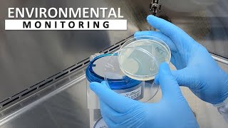
Microbial Monitoring: The systematic observation and analysis of microorganisms in water to assess safety and quality.
-
Culturing Method: A technique where microorganisms are grown in a nutrient medium to form visible colonies.
-
Flow Cytometry: A technology that analyzes and counts cells in a fluid as they pass through a detection apparatus.
-
Viability: The capability of microorganisms to grow and reproduce, crucial for assessing health risks.
Examples & Applications
Using a nutrient agar plate, researchers can plate a water sample and incubate it, leading to visible colonies that can be counted.
If 100 CFUs are identified in a diluted 1 ml sample, and it was diluted 10 times, this indicates 1,000 CFUs in the original 10 ml sample.
Memory Aids
Interactive tools to help you remember key concepts
Rhymes
Count the CFU with glee, in a colony they shall be, five per hundred, that’s our plea, CPCB’s number—easy as can be.
Stories
Once upon a time, a researcher ventured into the unclear waters, searching for hidden pathogens. With a microscope and a dream, they filtered, cultured, and finally saw the tiny colonies grow into flourishing cities on nutrient plates!
Memory Tools
Remember Counting Filters Under microscopes—C.F.U. for CFUs!
Acronyms
CWC - Count, Wash, Culture
the three key steps in microbial analysis.
Flash Cards
Glossary
- CPCB
Central Pollution Control Board, the regulatory authority that sets water quality standards in India.
- Microorganism
A microscopic organism, such as bacteria, fungi, viruses, and protozoa.
- Colony Forming Unit (CFU)
A unit used to estimate the number of viable microorganisms in a sample based on the number of colonies formed on a culture medium.
- Viable
Refers to living microorganisms that can grow and reproduce.
- Turbidity
The cloudiness or haziness of a fluid caused by large numbers of individual particles.
Reference links
Supplementary resources to enhance your learning experience.