
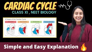
Cardiac Cycle
Enroll to start learning
You’ve not yet enrolled in this course. Please enroll for free to listen to audio lessons, classroom podcasts and take practice test.
Interactive Audio Lesson
Listen to a student-teacher conversation explaining the topic in a relatable way.
Introduction to the Cardiac Cycle
🔒 Unlock Audio Lesson
Sign up and enroll to listen to this audio lesson

Today, we are going to explore the cardiac cycle which is critical in our understanding of how the heart functions. Can anyone tell me what happens during the relaxation phase?

Isn't that when the blood fills the heart?
Exactly! This phase is called diastole, where the chambers are relaxed and blood flows in. It's essential for the heart's efficiency. Now, what do we call the phase when the heart contracts?

That would be systole!
Right again! During systole, the heart pumps blood out. Let's remember: 'D for Diastole is down, filling and relaxing; S for Systole is strong, pushing blood around.' Now, what can you conclude about the importance of these phases?

Both phases are necessary for effective blood circulation!
Great summary!
The Role of the Heart Valves
🔒 Unlock Audio Lesson
Sign up and enroll to listen to this audio lesson

Let's discuss the heart valves. Why do you think the valves are crucial during the cardiac cycle?

They probably prevent backflow of blood!
Exactly! The atrioventricular valves prevent backflow to the atria during ventricular contraction. Can anyone name those valves?
The tricuspid and bicuspid valves!
Correct! And what happens to the semilunar valves during ventricular systole?
They open to allow blood into the pulmonary artery and aorta.
That's right! Let’s use the mnemonic 'Saturate The Valve: S for Systole meaning open; D for Diastole meaning closed.' This can help remember their function related to contraction and relaxation.
The Heart's Electrical Activity
🔒 Unlock Audio Lesson
Sign up and enroll to listen to this audio lesson
The SAN is referred to as the pacemaker of the heart. Does anyone know why?
Because it generates action potentials that trigger heartbeats?
Exactly! The SAN generates electrical signals that start both atrial contraction and eventually lead to ventricular contraction via the AVN. Why do you think it's important for these events to be coordinated?
So that the heart can efficiently pump blood without mixing oxygenated and deoxygenated blood?
Spot on! Synchronism in contraction ensures efficient blood flow throughout the body. Remember: 'All points are right when SAN leads the fight!' This encapsulates the rhythm of heart activity.
Clinical Importance of the Cardiac Cycle
🔒 Unlock Audio Lesson
Sign up and enroll to listen to this audio lesson
Today, let’s discuss how understanding the cardiac cycle can aid in recognizing heart conditions. What recorded sound should we pay attention to?
The heart sounds, like 'lub' and 'dub'!
Correct! 'Lub' is associated with closure of the AV valves, while 'dub' relates to semilunar valve closure. Abnormalities in these sounds can indicate problems. How can we assess heart function?
Through an ECG?
Yes! An ECG is vital for monitoring electrical activities. Keep this in mind: 'Loud or Dub, heart's a club; with rhythms to share, diagnose with care!' This rhythmic framework for diagnosis is key!
Introduction & Overview
Read summaries of the section's main ideas at different levels of detail.
Quick Overview
Standard
The cardiac cycle describes the rhythmic process by which the heart functions, including the phases of contraction (systole) and relaxation (diastole) in the atria and ventricles, regulated primarily by the sino-atrial node. It accounts for the heart's pumping action and the mechanical movements that ensure proper blood flow.
Detailed
Detailed Summary of the Cardiac Cycle
The cardiac cycle is an essential physiological process that refers to the sequence of events involving the contraction (systole) and relaxation (diastole) of the heart's chambers. Each full cycle begins with all four chambers of the heart in a relaxed state, or joint diastole. During this period, blood flows passively from the pulmonary veins and vena cava into the left and right ventricles through open atrioventricular (tricuspid and bicuspid) valves, while the semilunar valves (guarding the pulmonary arteries and aorta) remain closed.
Once the sino-atrial node (SAN) generates an action potential, it initiates atrial contraction (atrial systole), pushing additional blood into the ventricles by about 30%. This electrical impulse spreads to the atrio-ventricular node (AVN) and down the bundle of His, causing the ventricles to contract (ventricular systole). This contraction elevates ventricular pressure, closing the atrioventricular valves to prevent backflow and opening the semilunar valves. Blood is then ejected into the pulmonary artery and aorta.
The ventricles then enter diastole as pressure decreases and the semilunar valves close. The cycle's duration averages 0.8 seconds with each ventricle pumping about 70 mL of blood per cycle (stroke volume). The total blood volume heart pumps in one minute (cardiac output) averages about 5 liters in a healthy adult. This cyclical contraction and relaxation, capable of producing heart sounds 'lub' and 'dub', is regulated by specialized pacemaker cells and can vary with physical activity levels.
Youtube Videos


Audio Book
Dive deep into the subject with an immersive audiobook experience.
Resting Phase of the Heart
Chapter 1 of 8
🔒 Unlock Audio Chapter
Sign up and enroll to access the full audio experience
Chapter Content
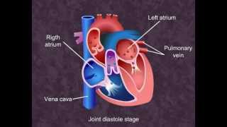
To begin with, all the four chambers of heart are in a relaxed state, i.e., they are in joint diastole. As the tricuspid and bicuspid valves are open, blood from the pulmonary veins and vena cava flows into the left and the right ventricle respectively through the left and right atria. The semilunar valves are closed at this stage.
Detailed Explanation
At the beginning of the cardiac cycle, all four chambers of the heart (the left atrium, left ventricle, right atrium, and right ventricle) are relaxed. This state is known as joint diastole, where the heart is not actively pumping. During this time, the tricuspid valve between the right atrium and right ventricle and the bicuspid valve (mitral valve) between the left atrium and left ventricle are open. Due to this opening, blood flows into the ventricles from the atria. The semilunar valves that lead to the arteries are closed, preventing backflow.
Examples & Analogies
Think of this phase like a calm waiting area in a train station, where travelers are seated and waiting for their train to arrive. Similarly, the heart is at rest, waiting for electrical impulses to trigger it to pump.
Atrial Contraction
Chapter 2 of 8
🔒 Unlock Audio Chapter
Sign up and enroll to access the full audio experience
Chapter Content
The SAN now generates an action potential which stimulates both the atria to undergo a simultaneous contraction – the atrial systole. This increases the flow of blood into the ventricles by about 30 per cent. The action potential is conducted to the ventricular side by the AVN and AV bundle.
Detailed Explanation
The Sinoatrial Node (SAN) acts as the heart's pacemaker. It generates an electrical impulse, which prompts both atria to contract at the same time, known as atrial systole. This contraction pushes additional blood (about 30%) into the ventricles, preparing them for the next phase. The electrical signal then travels through the Atrioventricular Node (AVN) and the AV bundle, setting up the next steps in the cycle.
Examples & Analogies
Imagine a synchronized dance performance where dancers (the atria) receive a cue from the conductor (the SAN). When the conductor gives the signal, all dancers move together, enhancing the performance, just like how blood flow into the ventricles is increased.
Ventricular Contraction
Chapter 3 of 8
🔒 Unlock Audio Chapter
Sign up and enroll to access the full audio experience
Chapter Content
This causes the ventricular muscles to contract (ventricular systole), the atria undergoes relaxation (diastole), coinciding with the ventricular systole. Ventricular systole increases the ventricular pressure causing the closure of tricuspid and bicuspid valves due to attempted backflow of blood into the atria.
Detailed Explanation
Once the atria have pushed blood into the ventricles, they relax again (diastole). Simultaneously, the ventricles contract (ventricular systole) due to the electrical signal from the AV bundle. This contraction increases the pressure in the ventricles, prompting the closure of the tricuspid and bicuspid valves to prevent the backflow of blood into the atria. This ensures that blood flows forward into the arteries instead.
Examples & Analogies
Think of a water balloon being filled (the ventricles). When you squeeze it (ventricular contraction), the only way for the water to go is out of the nozzle (the arteries), just as blood is propelled forward into circulation.
Ejection of Blood
Chapter 4 of 8
🔒 Unlock Audio Chapter
Sign up and enroll to access the full audio experience
Chapter Content
As the ventricular pressure increases further, the semilunar valves guarding the pulmonary artery (right side) and the aorta (left side) are forced open, allowing the blood in the ventricles to flow through these vessels into the circulatory pathways.
Detailed Explanation
When the pressure in the ventricles exceeds the pressure in the arteries, the semilunar valves open. This allows oxygen-poor blood to be ejected from the right ventricle into the pulmonary artery and oxygen-rich blood from the left ventricle into the aorta. This is a crucial step where blood is sent to the lungs for oxygenation and then to the rest of the body.
Examples & Analogies
Imagine a water slide at an amusement park; when the water builds up pressure enough to overcome the slide's top, it shoots down into the pool, just as blood is ejected into the circulatory system.
Relaxation Phase
Chapter 5 of 8
🔒 Unlock Audio Chapter
Sign up and enroll to access the full audio experience
Chapter Content
The ventricles now relax (ventricular diastole) and the ventricular pressure falls causing the closure of semilunar valves which prevents the backflow of blood into the ventricles. As the ventricular pressure declines further, the tricuspid and bicuspid valves are pushed open by the pressure in the atria exerted by the blood which was being emptied into them by the veins.
Detailed Explanation
After blood is ejected, the ventricles relax, entering a phase known as ventricular diastole. The pressure within the ventricles drops, leading to the closure of the semilunar valves to prevent backflow. As the ventricles continue to relax and pressure decreases, the blood returning from the body fills the atria, pushing open the tricuspid and bicuspid valves, allowing blood flow back into the ventricles.
Examples & Analogies
Think of a deflating balloon; as it loses air (pressure), it opens up, allowing more air to fill it. In this case, the relaxed ventricles receive more blood from the atria.
Completion of the Cardiac Cycle
Chapter 6 of 8
🔒 Unlock Audio Chapter
Sign up and enroll to access the full audio experience
Chapter Content
The ventricles and atria are now again in a relaxed (joint diastole) state, as earlier. Soon the SAN generates a new action potential and the events described above are repeated in that sequence and the process continues. This sequential event in the heart which is cyclically repeated is called the cardiac cycle.
Detailed Explanation
After the ventricles fill, the heart returns to the initial state of relaxation (joint diastole). As the SAN generates a new electrical impulse, the entire process starts over. This sequence of events that occurs with every heartbeat is known as the cardiac cycle.
Examples & Analogies
Consider the process of washing clothes in a machine. Once the wash cycle is done, it resets to start another wash. Similarly, the heart resets itself after completing a cycle to begin the next one.
Cardiac Cycle Duration and Output
Chapter 7 of 8
🔒 Unlock Audio Chapter
Sign up and enroll to access the full audio experience
Chapter Content
As mentioned earlier, the heart beats 72 times per minute, i.e., that many cardiac cycles are performed per minute. From this, it could be deduced that the duration of a cardiac cycle is 0.8 seconds. During a cardiac cycle, each ventricle pumps out approximately 70 mL of blood which is called the stroke volume.
Detailed Explanation
On average, the human heart beats around 72 times per minute. This helps us determine that the duration of an individual cardiac cycle is roughly 0.8 seconds. Each ventricle pumps about 70 mL of blood per cycle, known as stroke volume. This is crucial as it helps determine the efficiency of the heart and the blood volume circulating through the body.
Examples & Analogies
Imagine a water fountain. Each time it pumps out water (beat), it releases a specific amount (stroke volume). The frequency of the fountain's pumping helps us understand how much water is circulating into the air (cardiac cycles).
Understanding Heart Sounds
Chapter 8 of 8
🔒 Unlock Audio Chapter
Sign up and enroll to access the full audio experience
Chapter Content
During each cardiac cycle two prominent sounds are produced which can be easily heard through a stethoscope. The first heart sound (lub) is associated with the closure of the tricuspid and bicuspid valves whereas the second heart sound (dub) is associated with the closure of the semilunar valves.
Detailed Explanation
As the heart goes through its cycles, two main sounds can be heard: 'lub' and 'dub'. The 'lub' sound occurs when the tricuspid and bicuspid valves close after ventricular contraction. The 'dub' sound is made when the semilunar valves close after the blood has been pumped into the arteries. These sounds are important indicators of heart function and can be used to diagnose potential issues.
Examples & Analogies
Think of the heart sounds as the sounds of a drum in a musical performance. The 'lub' is like a strong bass drum beat marking a key moment, while the 'dub' is a secondary drum that adds rhythm to the performance, helping to signal the end of a cycle.
Key Concepts
-
Cardiac Cycle: Involves systole and diastole, crucial for heart function.
-
Systole and Diastole: Systole is contraction, diastole is relaxation of heart chambers.
-
Role of SAN: The sino-atrial node regulates heart rhythm and initiates contraction.
Examples & Applications
During each cardiac cycle, approximately 70 mL of blood is pumped from each ventricle.
The heart sounds 'lub' and 'dub' are produced by the closure of valves during the cardiac cycle.
Memory Aids
Interactive tools to help you remember key concepts
Rhymes
'Lub' is the sound of valves that shut, 'Dub' means the semilunars do their cut.
Stories
Imagine a race where the SAN is the starter's gun, signaling the atria at the beginning and the ventricles to run!
Memory Tools
Remember ADS for Atrial Diastole followed by Systole.
Acronyms
SAD
Systole Atria
Diastole relaxing phase.
Flash Cards
Glossary
- Cardiac Cycle
The sequence of events during one heartbeat, including the contraction and relaxation of heart chambers.
- Systole
The phase of the cardiac cycle where the heart contracts to pump blood.
- Diastole
The phase of the cardiac cycle where the heart relaxes and fills with blood.
- Sinoatrial Node (SAN)
The heart's natural pacemaker that generates electrical impulses for heart contractions.
- Atrioventricular Node (AVN)
A cluster of cells that receive impulses from the SAN and transmit them to the ventricles.
- Stroke Volume
The volume of blood pumped from each ventricle per heartbeat.
- Cardiac Output
The total volume of blood pumped by the heart per minute.
Reference links
Supplementary resources to enhance your learning experience.