
Neuron as Structural and Functional Unit of Neural System
Enroll to start learning
You’ve not yet enrolled in this course. Please enroll for free to listen to audio lessons, classroom podcasts and take practice test.
Interactive Audio Lesson
Listen to a student-teacher conversation explaining the topic in a relatable way.
Structure of a Neuron
🔒 Unlock Audio Lesson
Sign up and enroll to listen to this audio lesson

Today we will dive into the structure of neurons, which are crucial to the functioning of our neural system. Can anyone tell me the main parts of a neuron?

Is it the cell body, dendrites, and axon?
Exactly! The cell body contains essential organelles, the dendrites receive signals, and the axon transmits impulses. Remember this using the acronym CDA: C for Cell body, D for Dendrites, and A for Axon.

Why are dendrites important?
Dendrites are like antennas that receive signals from other neurons, ensuring that the neuron can communicate effectively. Now, how do you think axons work?

They send messages away from the cell body!
Correct! The axon can be myelinated or unmyelinated, impacting the speed of signal transmission. Let's remember myelinated with the phrase 'Myelin is Mighty' because it speeds up conduction.

What about the Nissl’s granules?
Good question! Nissl's granules are involved in protein synthesis within the neuron. In summary, the three parts of a neuron work together to receive and transmit signals efficiently.
Mechanism of Nerve Impulse Generation
🔒 Unlock Audio Lesson
Sign up and enroll to listen to this audio lesson

Now that we know the structure, let's discuss how nerve impulses are generated. Who can explain what resting potential is?
It's the state when the neuron is not sending signals, right?
Exactly! During resting potential, there's a difference in ion concentrations inside and outside the neuron, primarily potassium and sodium ions. Remember, K+ is 'In' while Na+ is 'Out'.
What happens when a stimulus occurs?
Great question! When a stimulus triggers the neuron, sodium channels open, causing depolarisation, and this shift leads to an action potential. Think of it as a wave: depolarisation spreads along the axon.
So, the action potential propagates along the axon?
Yes! It creates a current flow that continues the process, ensuring rapid impulse transmission. Always remember: 'Depolarize to Propagate!'
What about the recovery phase?
After depolarisation, the neuron repolarizes, restoring resting potential, making it responsive again. This entire process forms the basis of how our nervous system communicates.
Transmission of Nerve Impulses at Synapses
🔒 Unlock Audio Lesson
Sign up and enroll to listen to this audio lesson
Let's move on to how impulses transmit from one neuron to another at synapses. What types of synapses can you name?
There's electrical and chemical synapse!
Right! Electrical synapses allow direct current flow, while chemical synapses use neurotransmitters. Think of neurotransmitters as 'The Messengers of the Brain.'
Can you explain how chemical synapses work?
Of course! When the action potential reaches the axon terminal, synaptic vesicles containing neurotransmitters fuse with the membrane and release them into the synaptic cleft!
And then they bind to receptors on the next neuron?
Exactly! That binding opens ion channels in the postsynaptic neuron, potentially generating a new action potential. Remember: 'Release to Receive!'
What if the neurotransmitter doesn't lead to an action potential?
Good follow-up! Some neurotransmitters can inhibit impulses. Hence, neurotransmitters can be excitatory or inhibitory, altering neuron activity depending on the situation. The balance is critical for proper function!
Introduction & Overview
Read summaries of the section's main ideas at different levels of detail.
Quick Overview
Standard
Neurons consist of three main parts: the cell body, dendrites, and axon. The section discusses their roles in transmitting nerve impulses, the mechanisms of resting and action potentials, and the process of impulse transmission across synapses.
Detailed
Neuron as Structural and Functional Unit of Neural System
Neurons are the basic structural and functional units of the neural system, enabling the rapid transmission of signals throughout the body. Each neuron comprises three primary components: the cell body, which contains organelles including Nissl’s granules; the dendrites, which receive signals from other neurons; and the axon, responsible for transmitting impulses away from the cell body. Axons are typically long and may be myelinated or unmyelinated depending on the presence of a myelin sheath, which increases conduction efficiency. Myelinated fibers are found in spinal and cranial nerves, while unmyelinated fibers are often in the autonomic nervous system.
Key Functions of Neurons
The polarisation of the neuron’s membrane is essential for maintaining the resting potential, which is crucial for nerve impulse generation. When triggered, the neuron experiences depolarisation, resulting in an action potential that creates a wave of depolarisation and repolarisation along the axon.
Impulse Transmission
Neurons communicate via synapses, with two main types being electrical and chemical synapses. Electrical synapses allow direct current flow between neurons, while chemical synapses involve the release of neurotransmitters from the presynaptic neuron to the postsynaptic neuron through the synaptic cleft. This section underscores the significance of neurons in coordinating rapid signals essential for body functions.
Youtube Videos


Audio Book
Dive deep into the subject with an immersive audiobook experience.
Basic Structure of a Neuron
Chapter 1 of 5
🔒 Unlock Audio Chapter
Sign up and enroll to access the full audio experience
Chapter Content
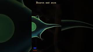
A neuron is a microscopic structure composed of three major parts, namely, cell body, dendrites and axon. The cell body contains cytoplasm with typical cell organelles and certain granular bodies called Nissl’s granules. Short fibres which branch repeatedly and project out of the cell body also contain Nissl’s granules and are called dendrites. These fibres transmit impulses towards the cell body. The axon is a long fibre, the distal end of which is branched. Each branch terminates as a bulb-like structure called synaptic knob which possess synaptic vesicles containing chemicals called neurotransmitters. The axons transmit nerve impulses away from the cell body to a synapse or to a neuro-muscular junction.
Detailed Explanation
A neuron is essentially the basic building block of the nervous system. It has three key components:
- Cell Body: This is the main part of the neuron where the nucleus and organelles are located. It supports the metabolic activities of the neuron.
- Dendrites: These are short, branched extensions that receive signals from other neurons and transmit them to the cell body.
- Axon: This long fiber transmits impulses away from the cell body towards other neurons or muscles. The end of the axon has structures called synaptic knobs that release neurotransmitters to facilitate communication between neurons.
Understanding these parts is crucial as they illustrate how neurons communicate and process information.
Examples & Analogies
Think of a neuron like a communication network in a company. The cell body is akin to the headquarters, where decisions are made. Dendrites are like the email systems that receive messages from various branches (other neurons). The axon is like the postal service that sends out information and instructions to different branches and customers (other neurons or muscles) to maintain the company's operations.
Types of Neurons
Chapter 2 of 5
🔒 Unlock Audio Chapter
Sign up and enroll to access the full audio experience
Chapter Content
Based on the number of axon and dendrites, the neurons are divided into three types, i.e., multipolar (with one axon and two or more dendrites; found in the cerebral cortex), bipolar (with one axon and one dendrite, found in the retina of eye) and unipolar (cell body with one axon only; found usually in the embryonic stage).
Detailed Explanation
Neurons can be classified based on their structure into three main types:
- Multipolar Neurons: These neurons have one axon and multiple dendrites, allowing them to connect with many other neurons. They are primarily found in the brain's cerebral cortex, where complex processing occurs.
- Bipolar Neurons: These have one axon and one dendrite. They are less common and are mainly found in specific sensory organs, such as the retina of the eye, where they help transmit visual information.
- Unipolar Neurons: These consist of a single axon that branches into two directions. They are primarily found in the early stages of development (embryonic stage) and are involved in sensory information processing.
This classification helps us understand how different neurons are adapted for specific functions.
Examples & Analogies
Imagine the types of phones in a large office. Multipolar neurons are like smartphones, capable of handling multiple calls and notifications at once (many connections). Bipolar neurons can be compared to a traditional phone that can only make or receive one call at a time (one connection). Unipolar neurons are like emergency phones that connect directly to one point, typically used in critical situations (very specific functions).
Axon Types and Myelination
Chapter 3 of 5
🔒 Unlock Audio Chapter
Sign up and enroll to access the full audio experience
Chapter Content
There are two types of axons, namely, myelinated and non-myelinated. The myelinated nerve fibres are enveloped with Schwann cells, which form a myelin sheath around the axon. The gaps between two adjacent myelin sheaths are called nodes of Ranvier. Myelinated nerve fibres are found in spinal and cranial nerves. Unmyelinated nerve fibre is enclosed by a Schwann cell that does not form a myelin sheath around the axon, and is commonly found in autonomous and the somatic neural systems.
Detailed Explanation
Axons can be either myelinated or unmyelinated:
- Myelinated Axons: These axons are covered with a fatty layer called the myelin sheath, produced by Schwann cells. The myelin sheath acts as insulation, which speeds up the transmission of nerve impulses along the axon. The myelin sheath is not continuous; it is interrupted at intervals by small gaps known as nodes of Ranvier, where the axonal membrane is exposed. This arrangement allows for rapid impulse conduction via saltatory conduction, where the impulse jumps from node to node.
- Unmyelinated Axons: In contrast, unmyelinated axons lack this insulating layer, resulting in slower impulse transmission. These are found in areas of the autonomic and somatic nervous systems where quick response is less critical.
Understanding the difference between myelinated and unmyelinated axons is crucial because it impacts how quickly our bodies can respond to stimuli.
Examples & Analogies
Think of myelinated axons like cars driving on a highway with express lanes (myelinated), allowing them to travel faster, especially when changing lanes at exits (nodes of Ranvier). Unmyelinated axons, on the other hand, are like cars on a city street: they move slower because they face more stops (no myelin) and have to navigate through traffic signals (impulse transmission happens more gradually).
Generation and Conduction of Nerve Impulse
Chapter 4 of 5
🔒 Unlock Audio Chapter
Sign up and enroll to access the full audio experience
Chapter Content
Neurons are excitable cells because their membranes are in a polarised state. The electrical potential difference across the resting plasma membrane is called the resting potential. When a stimulus is applied at a site on the polarised membrane, the membrane at that site becomes freely permeable to Na+. This leads to a rapid influx of Na+ followed by the reversal of the polarity at that site, i.e., the outer surface of the membrane becomes negatively charged and the inner side becomes positively charged. This process is known as depolarisation.
Detailed Explanation
The generation and conduction of a nerve impulse involve several steps:
1. Polarised State: Neurons maintain a resting potential due to concentrations of ions, primarily sodium (Na+) and potassium (K+). That means the inside of the neuron is negative compared to the outside.
2. Stimulus Application: When a stimulus occurs, it makes the neuron's membrane at that specific site become permeable to sodium ions (Na+).
3. Depolarisation: As Na+ rushes into the neuron, the internal charge shifts to positive. This change in charge is called depolarisation and creates an action potential.
4. Impulse Conduction: Once depolarised, the action potential triggers further depolarisations down the axon, allowing the nerve impulse to travel rapidly.
Understanding this process is crucial for grasping how signals travel through our nervous system.
Examples & Analogies
Think of this process like a line of dominos. The initial domino (the stimulus) falls over (depolarisation) and causes the next domino to tip (impulse conduction) and so on down the line. Just as the falling domino sets off a chain reaction, the initial change in the neuron's charge leads to an impulse that rapidly travels along the neuron.
Transmission of Impulses Across Synapses
Chapter 5 of 5
🔒 Unlock Audio Chapter
Sign up and enroll to access the full audio experience
Chapter Content
A nerve impulse is transmitted from one neuron to another through junctions called synapses. A synapse is formed by the membranes of a pre-synaptic neuron and a post-synaptic neuron, which may or may not be separated by a gap called synaptic cleft. There are two types of synapses, namely, electrical synapses and chemical synapses. At electrical synapses, the membranes of pre- and post-synaptic neurons are in very close proximity.
Detailed Explanation
Synapses are critical for communication between neurons:
1. Synapse Structure: A synapse connects a pre-synaptic neuron (sending neuron) and a post-synaptic neuron (receiving neuron). There may be a small gap between them called the synaptic cleft.
2. Types of Synapses:
- Electrical Synapses: These allow direct flow of electrical signals between neurons. The membranes are very close, permitting rapid transmission of impulses. They function similarly to impulse conduction along an axon. These are typically faster but less common.
- Chemical Synapses: At these synapses, the neurons do not touch. Instead, the pre-synaptic neuron releases neurotransmitters into the synaptic cleft. These chemicals then bind with receptors on the post-synaptic neuron, generating a new potential that can lead to an action potential.
Understanding how impulses are transmitted across synapses is essential, as it explains how signals propagate through complex neural networks.
Examples & Analogies
Imagine synapses as two people passing a message through a wall. Electrical synapses are like passing a message through a hole in the wall (quick and direct), while chemical synapses are like writing the message down on a piece of paper (neurotransmitter) and handing it over. The second person reads the message and acts upon it (new potential) on their side.
Key Concepts
-
Neuron: The basic unit of the neural system responsible for transmitting impulses.
-
Dendrites: Structures that receive signals from other neurons.
-
Axon: The component that transmits the signal away from the neuron.
-
Resting Potential: The negative charge maintained across the neuron membrane at rest, necessary for action potentials.
-
Myelination: The process of wrapping axons in a myelin sheath, which increases conduction speed.
-
Synapse: The junction where neurons communicate with each other.
Examples & Applications
A multipolar neuron is typically found in the cerebral cortex and has multiple dendrites and one axon.
The process of action potential begins with depolarisation at the axon membrane when a stimulus is applied.
Chemical synapses require the release of neurotransmitters to transmit impulses, while electrical synapses allow direct current flow.
Memory Aids
Interactive tools to help you remember key concepts
Rhymes
Dendrites receive, axons send, together they help signals blend.
Stories
Imagine a mailman (axon) delivering messages (nerve impulses) sent from homes (dendrites) to the main office (cell body).
Memory Tools
Remember CDA for the parts of a neuron: C for Cell body, D for Dendrites, A for Axon.
Acronyms
MINE
Myelination Increases Neural Efficiency.
Flash Cards
Glossary
- Neuron
Specialized cell that can transmit nerve impulses.
- Dendrites
Branch-like structures on neurons that receive messages.
- Axon
Long fiber that transmits impulses away from the neuron.
- Resting Potential
The electrical potential difference across a neuron’s membrane at rest.
- Action Potential
A rapid rise and fall in membrane potential that constitutes a nerve impulse.
- Synapse
The junction between two neurons, where impulse transmission occurs.
- Neurotransmitters
Chemicals that transmit signals across synapses.
- Myelination
The process of forming a myelin sheath around nerve fibers.
Reference links
Supplementary resources to enhance your learning experience.