
Structural Characterization
Interactive Audio Lesson
Listen to a student-teacher conversation explaining the topic in a relatable way.
Introduction to X-ray Diffraction (XRD)
🔒 Unlock Audio Lesson
Sign up and enroll to listen to this audio lesson

Today we will discuss X-ray Diffraction, a powerful technique for analyzing crystal structures. Does anyone know what Bragg's law states?

Is it something like nλ = 2d sinθ? That’s what I remember.
Exactly! Bragg's law describes the condition for constructive interference of scattered X-rays. This law helps us calculate lattice constants from the diffraction patterns.

How does knowing the lattice constant help us in semiconductor characterization?
Great question! It helps in determining the material's quality and detecting any strain or defects present, which can affect device performance.
Understanding SEM
🔒 Unlock Audio Lesson
Sign up and enroll to listen to this audio lesson

Now, let's shift our focus to Scanning Electron Microscopy, or SEM. Can anyone explain what makes SEM valuable for semiconductor characterization?

I think it gives us detailed images of the surface at a very high resolution.
That's right! SEM can analyze surfaces at the nanometer scale, allowing us to evaluate critical dimensions accurately. Can someone give me an application for SEM?

It can be used to observe the morphology of materials, right?
Absolutely! SEM's ability to combine imaging with Energy-Dispersive X-ray Spectroscopy enhances our understanding of composition as well.
Combining XRD and SEM
🔒 Unlock Audio Lesson
Sign up and enroll to listen to this audio lesson
Let’s discuss how XRD and SEM complement each other in structural characterization. Why might we want to use both?
Using both would give us a full picture of the material's structure and surface.
Exactly! XRD provides information about the crystal structure and lattice parameters, whereas SEM gives insights into the surface morphology and dimensional accuracy. Together, they validate our fabrication processes.
Can defects be detected through these methods?
Yes! While XRD shows strain and disturbances in the crystal structure, SEM can visually reveal surface defects. This combined data is crucial for optimizing semiconductor performance.
Introduction & Overview
Read summaries of the section's main ideas at different levels of detail.
Quick Overview
Standard
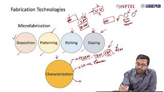
This section details two primary techniques for structural characterization: X-ray Diffraction (XRD) used for crystal structure analysis and Scanning Electron Microscopy (SEM) for examining surface morphology and dimensional accuracy. These methods are crucial for understanding the material properties and validating fabrication processes in semiconductor devices.
Detailed
Structural Characterization
Structural characterization is essential for understanding semiconductor materials and devices, focusing predominantly on their crystal structure and surface properties. This section discusses two pivotal techniques used in this area:
1. X-ray Diffraction (XRD)
XRD is employed to analyze the crystal structure of semiconductor materials. By applying Bragg's law (nλ = 2d sinθ), XRD helps researchers determine the lattice constants and the presence of any potential strain within the material.
2. Scanning Electron Microscopy (SEM)
SEM enables high-resolution imaging of surface morphology and critical dimensions of semiconductor materials. In addition to imaging, SEM can be coupled with Energy-Dispersive X-ray Spectroscopy (EDS) for compositional analysis, identifying different elements in the material. Both techniques are pivotal in material verification and play significant roles in the fabrication and evaluation of semiconductor devices.
Youtube Videos
Audio Book
Dive deep into the subject with an immersive audiobook experience.
X-ray Diffraction (XRD)
Chapter 1 of 2
🔒 Unlock Audio Chapter
Sign up and enroll to access the full audio experience
Chapter Content
3.4.1 X-ray Diffraction (XRD)
- Crystal structure analysis
- Lattice constant calculation (Bragg's law: nλ = 2d sinθ)
- Strain measurement
Detailed Explanation
X-ray Diffraction (XRD) is a technique used to analyze the crystal structure of materials. When X-rays hit a crystal, they are scattered in specific directions. By measuring these angles, we can determine the arrangement of atoms in the material, known as its crystal structure. The lattice constant, which describes the size of the unit cell in the crystal structure, can be calculated using Bragg's law, which explains the relationship between the wavelength of the X-ray (λ), the angle of diffraction (θ), and the distance between crystal planes (d). Additionally, XRD can also measure strain in the material, which indicates any distortions in the crystal lattice.
Examples & Analogies
Think of XRD like a detailed map of a city. Just as a map shows how streets are arranged and connected, XRD reveals how atoms are arranged in a solid. If you imagine a city experiencing construction or changes (strain), a map can help identify where those changes occur—similar to how XRD identifies strain in a crystal.
Scanning Electron Microscopy (SEM)
Chapter 2 of 2
🔒 Unlock Audio Chapter
Sign up and enroll to access the full audio experience
Chapter Content
3.4.2 Scanning Electron Microscopy (SEM)
- Surface morphology
- Critical dimension measurement
- Energy-dispersive X-ray spectroscopy (EDS) for composition
Detailed Explanation
Scanning Electron Microscopy (SEM) is a powerful technique used to observe the surface morphology (shape and structure) of a sample at much higher resolutions than optical microscopes. In SEM, a focused beam of electrons scans across the sample surface. The interaction between the electrons and the sample provides detailed images of the surface features. It can measure critical dimensions, such as widths and heights of structures with great precision. Additionally, SEM can be equipped with Energy-dispersive X-ray Spectroscopy (EDS), an analytical technique that helps identify the elemental composition of the sample by detecting X-rays emitted from the sample during electron bombardment.
Examples & Analogies
Imagine you're looking at a beach using a normal pair of binoculars, which can only show you a broad view. Now, think of SEM as a high-power microscope that lets you explore tiny grains of sand on that beach up close, revealing details about their texture and composition. Just like how a tool can assess the quality of grains and their arrangement on the beach, SEM does the same for the materials we study in the lab.
Key Concepts
-
X-ray Diffraction: A technique for studying crystal structures using X-rays.
-
Scanning Electron Microscopy: A method for visualizing the surface morphology of materials.
-
Bragg's Law: The relationship governing the diffraction of X-rays by crystalline materials.
Examples & Applications
Using XRD to determine the lattice constant of a silicon crystal, revealing its quality and impurities.
Implementing SEM to analyze the surface coatings on semiconductor materials and detect any defects.
Memory Aids
Interactive tools to help you remember key concepts
Rhymes
When X-ray beams dance just right, Crystal structures come to light.
Stories
Imagine you’re a detective investigating a crystal. XRD reveals its hidden layers, while SEM captures its smooth surface, helping you understand its story.
Memory Tools
Remember XRD as 'We Read the Diffraction' to remember it’s about crystal structure.
Acronyms
SEM= See Every Micron; a reminder that the technique captures high-resolution images.
Flash Cards
Glossary
- Xray Diffraction (XRD)
A technique used to determine the crystal structure and lattice constants of materials through the diffraction of X-rays.
- Bragg's Law
A fundamental equation used in XRD to relate the wavelength of X-rays to the angle of diffraction and the lattice spacing.
- Scanning Electron Microscopy (SEM)
A type of electron microscope that provides high-resolution images of a sample's surface morphology.
- EnergyDispersive Xray Spectroscopy (EDS)
An analytical technique that can be attached to SEM to identify the elemental composition of materials.
Reference links
Supplementary resources to enhance your learning experience.